There are some discussions related with the therapeutic group today, so I'll just post here for clarity:
-
Continue to verify Ab binding (as what the therpeutic group is doing)
-
Assess whether Beva and NRP1 can simultaneously bind to VEGF in a non-mutuall exclusive way: --> Coat the plate with Beva --> Add VEGF --> Wash off unbound VEGF --> Add NRP1 and detect NRP1 binding
if both can bind, it suggests that their binding sites on VEGF are distinct or not mutually exclusive.
- When time allows, also try using an HPD-containing NRP1-binding motif from VEGF165. (VEGF121 without HPD binds NRP1 with low affinity, but VEGF165 with HPD can binds with high affinity, so still use HPD for NRP1 binding and cell internalization).
The idea is that (i took a reference from Pinetree patent (https://patents.google.com/patent/WO2024030341A1/en):
EGFR binder brings the molecule to EGFR on the tumor cell surface → NRP1 binder grabs NRP1 on the same cell → EGFR and NRP1 are pulled close together → the EGFR–bispecific–NRP1 complex gets internalized into the cell → the complex is sent to the lysosome → EGFR, and possibly NRP1, are degraded (EGFR lysosome degradation is validated by lysosomal degradation inhibitor/proteasome inhibitor experiment) → less EGFR remains on the cell surface → EGFR signaling goes down → EGFR-dependent tumor cell growth/survival decrease
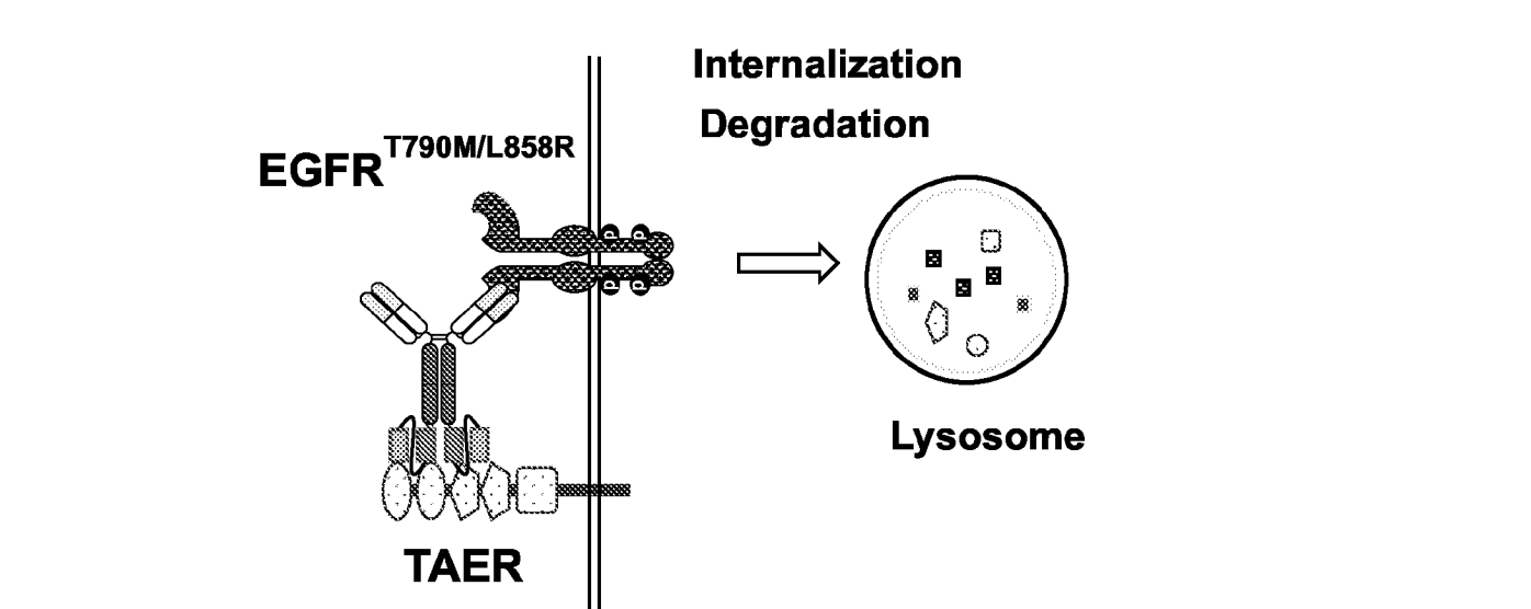
So in our case, the question is whether we can use an NRP1-binding HPD motif together with an EGFR-binding arm to help drag EGFR into the NRP1-mediated internalization/lysosomal degradation route, rather than only blocking EGFR signaling.
- VEGF daizy chaining in the Fab arm, and the buttom scFv like EGFR and MET (like #13), figure out cell activity (figure out explanation in either direction)
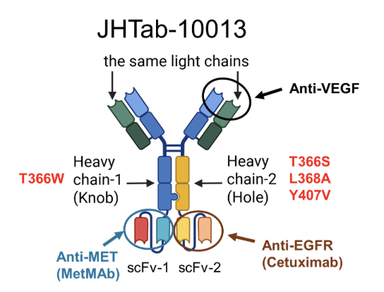
Anti-VEGF double arms bind soluble VEGF → VEGF helps bring multiple antibody molecules close together → a VEGF-dependent daisy-chain / Ab cluster forms → the antibody complex becomes enriched in the VEGF-rich tumor microenvironment → the EGFR binder grabs EGFR on the tumor cell surface → the MET binder grabs MET on the same cell → EGFR and MET are pulled close together in one antibody complex → EGFR and MET are occupied, so EGF/HGF-driven activation is blocked → the EGFR-bispecific-MET complex gets internalized into the cell → the complex is sent to the lysosome → EGFR and MET are downregulated / degraded → less EGFR and MET remain on the cell surface → EGFR/MET signaling goes down
Please feel free to correct me if my understanding is wrong.
Have a great weekend!
Best,
Lily